sel otak jika dilihat dari microscop
 04.51
04.51
 dani ahmad mubarok

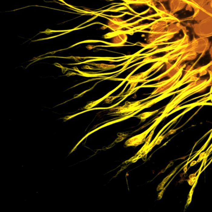
Photomicrograph of a neuron’s cell body (top, center) and its dendrites radiating out of it, obtained with a scanning electron microscope.
Photomicrograph of the molecular scaffolding of axons.

Photomicrograph of different components of the rat cerebellum, including Purkinje neurons in green, glia (non-neuronal cells) in red, and cell nuclei in blue.

sumber :http://www.amusingplanet.com/2010/10/portraits-of-mind-brain-cells-under.html


0 Response to "sel otak jika dilihat dari microscop"
Posting Komentar